Adnan
July 2026
Thank you,Dr Koh, for the excellent dental implant treatment.The whole process was well explained,smooth and comfortable.Im very pleased with the results and would definitely recommend this clinic to others
View on Google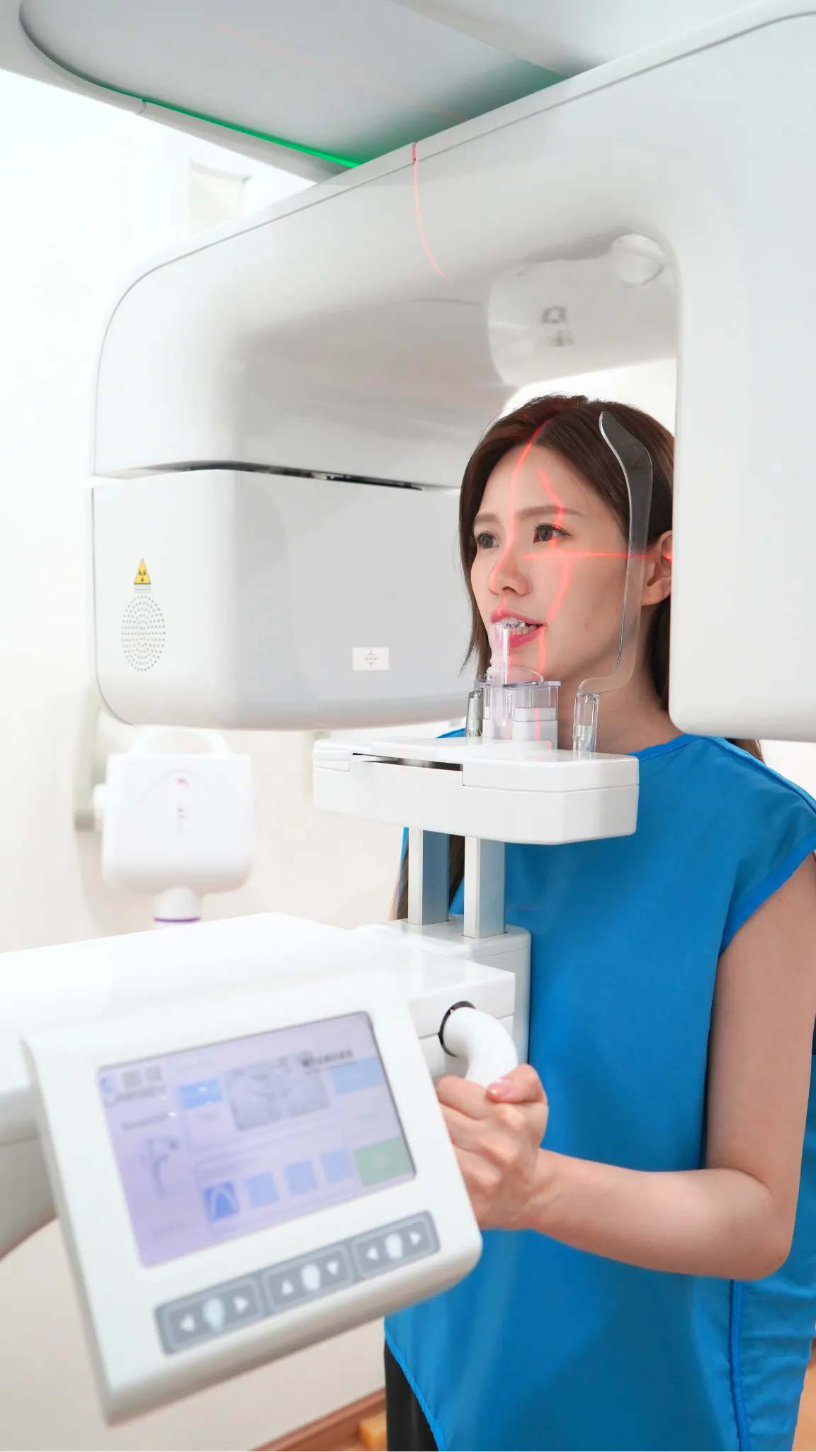
Digital periapical X-rays, OPG, lateral cephalometric X-rays and 3D CBCT scans taken in the clinic, only when they change your diagnosis or plan.
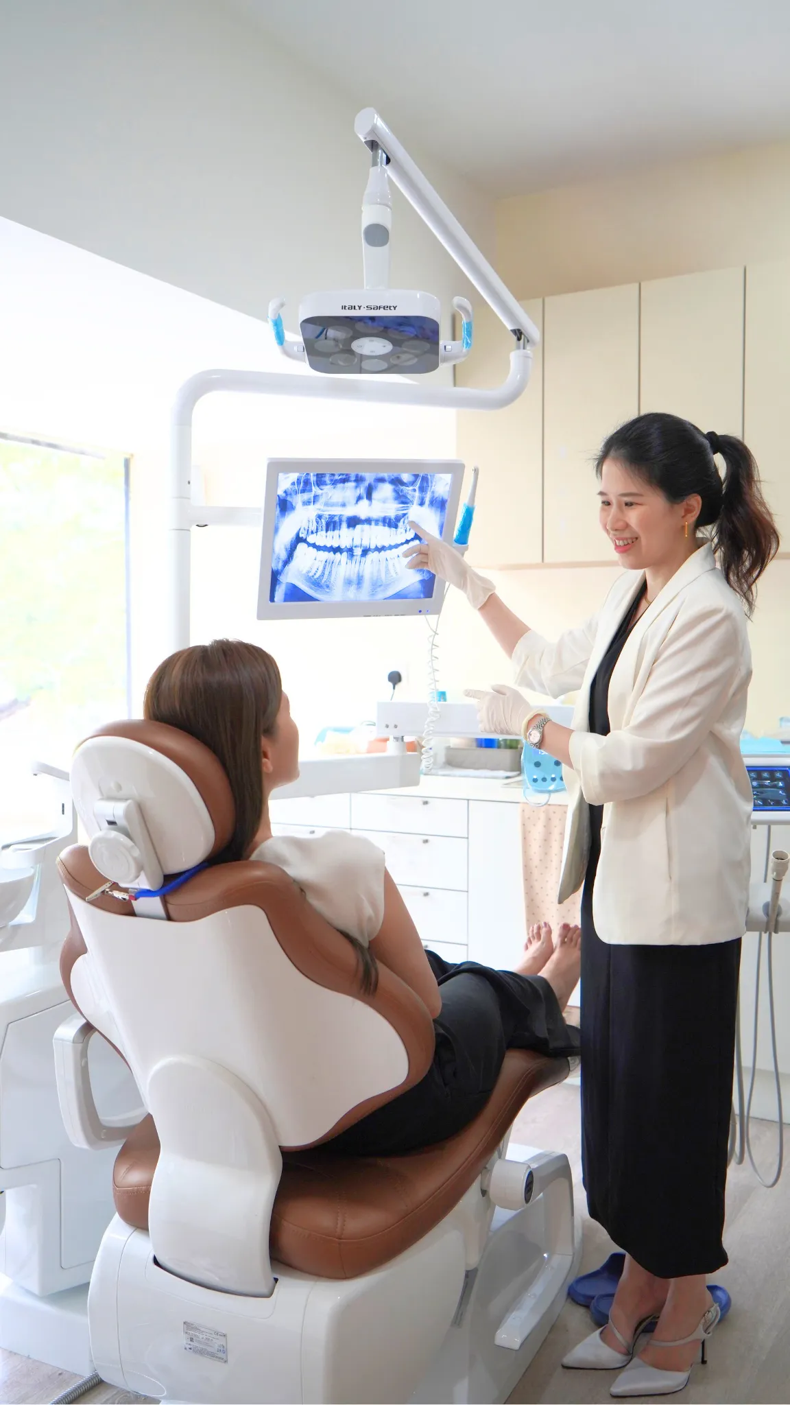
Dental X-rays show what cannot be seen by looking in the mouth: decay between teeth, infection at the root tip, bone levels around the teeth, the position of wisdom teeth and the shape of the jaw. We use small periapical X-rays for individual teeth, a panoramic OPG for the whole mouth, a lateral cephalometric X-ray for orthodontic planning, and a 3D CBCT scan for implants, surgery and complex root canal cases. All are digital, which keeps the radiation dose low and the images available immediately.
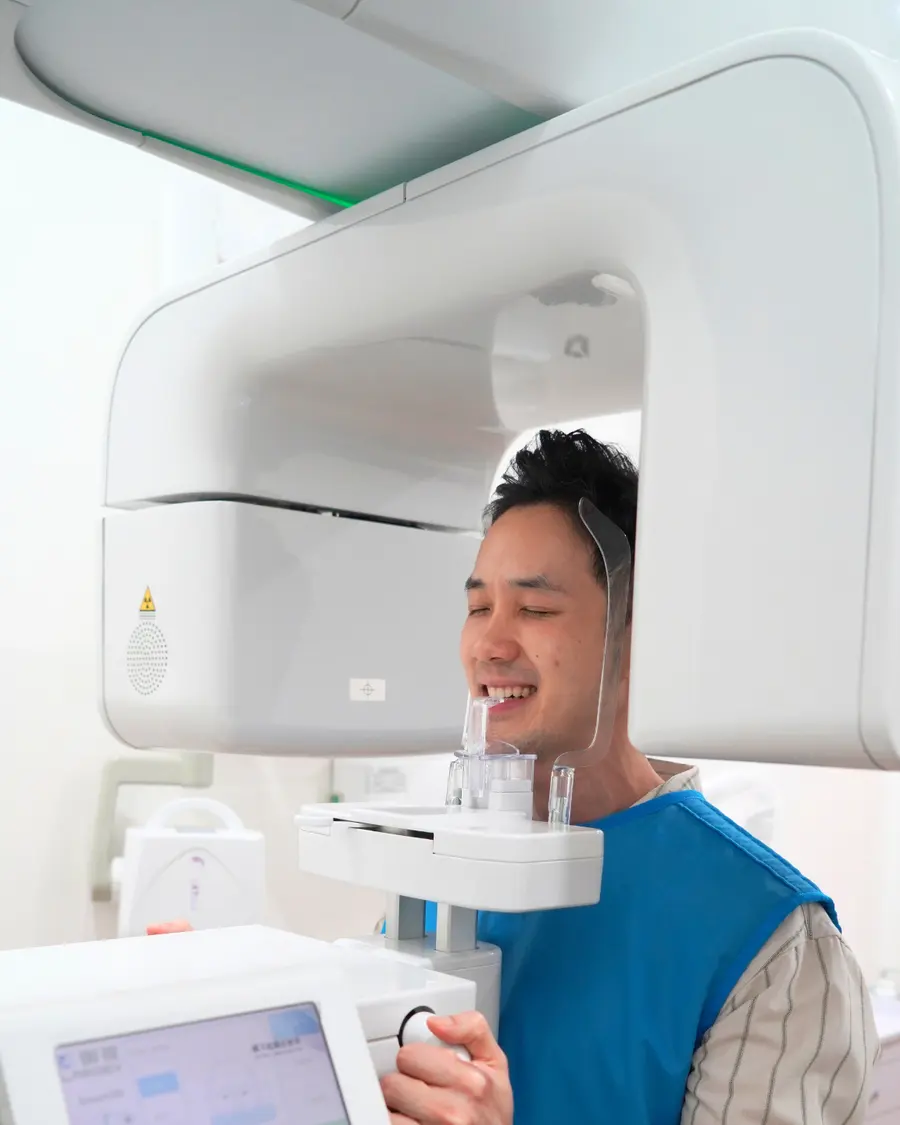
An intraoral X-ray is comparable to a day or two of background radiation.
Images appear on screen immediately.
No routine exposures without a reason.
Each image answers a specific question about your teeth or jaw.
Not every option suits every patient. We confirm the right one at your examination and explain it before anything starts.
Explore the options. The plan is tailored to your case.
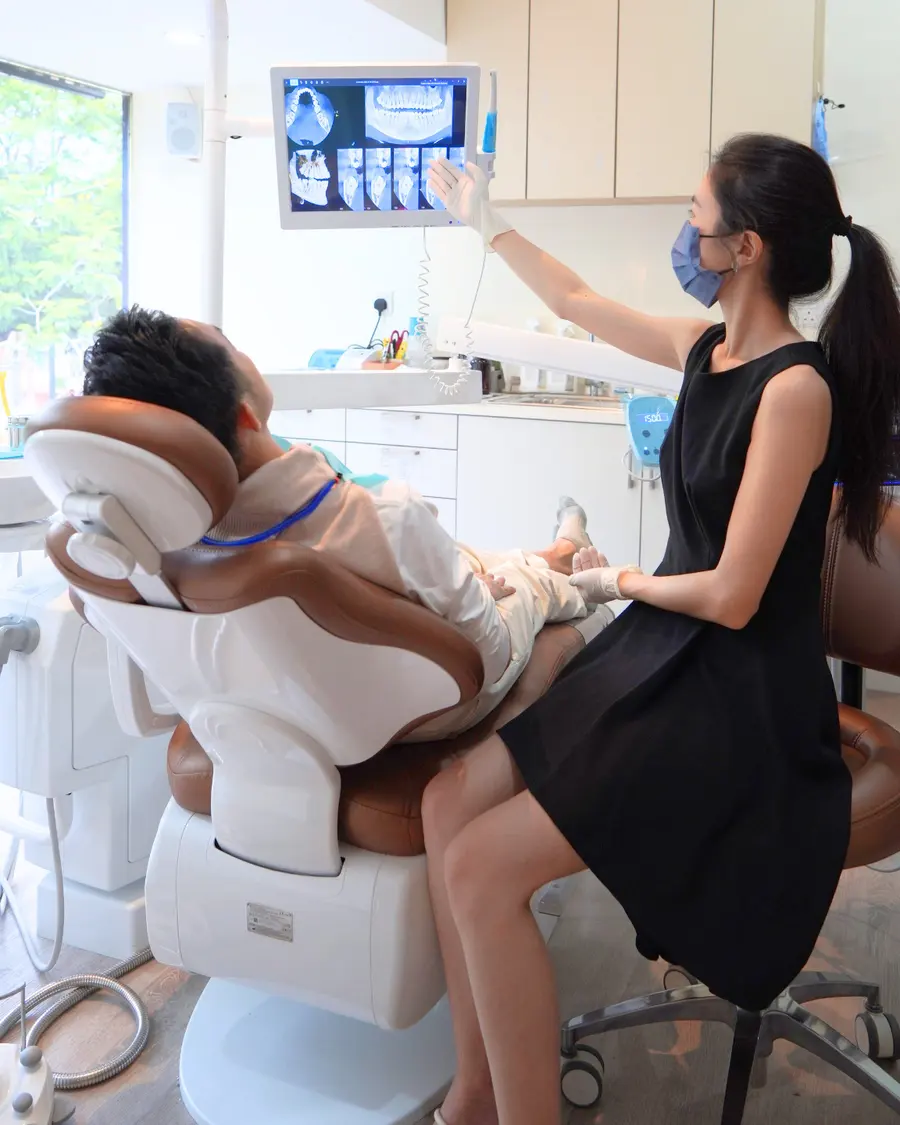
A Periapical (PA) X-ray provides a detailed image of an individual tooth, including the root and surrounding bone structure. It helps diagnose infections, decay, bone loss, and root-related problems with high accuracy. This quick and low-radiation imaging method is commonly used for root canal treatment and dental evaluations.
An OPG (Orthopantomogram) X-ray provides a panoramic view of the entire mouth, including all teeth, jawbones, and surrounding structures. It is commonly used for wisdom tooth evaluation, orthodontic treatment, dental implants, and comprehensive oral examinations.
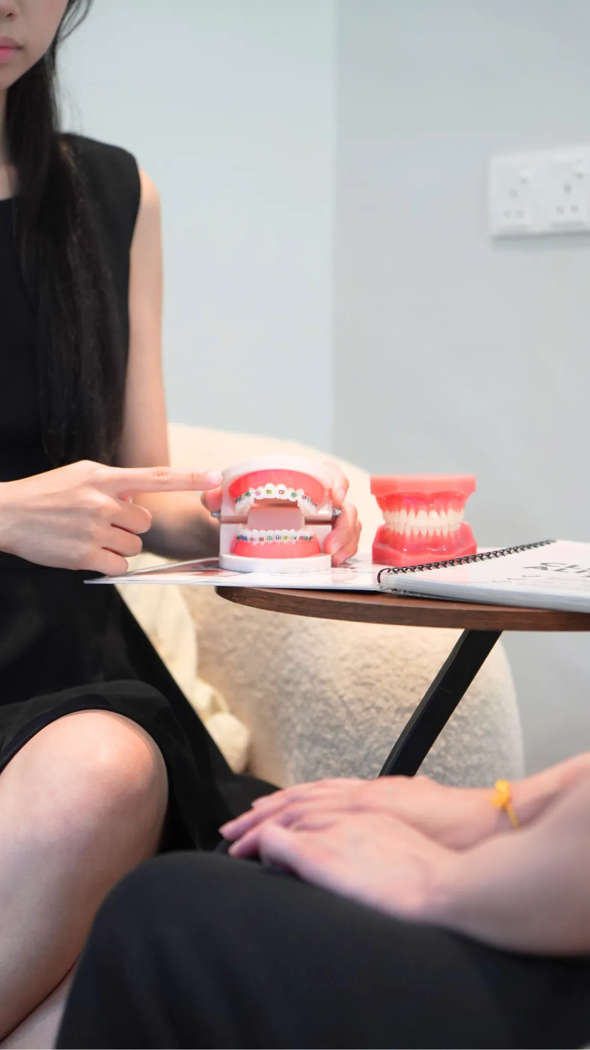
A Lateral Cephalometric X-ray is a side-profile dental imaging technique commonly used in orthodontics. It helps dentists and orthodontists evaluate jaw alignment, facial structure, and bite relationships for braces and corrective treatment planning.
A CBCT (Cone Beam Computed Tomography) scan provides advanced 3D dental imaging for precise diagnosis and treatment planning. It allows dentists to evaluate bone structure, nerves, teeth, and surrounding anatomy with high accuracy, making it useful for implants, surgery, and complex dental procedures.
What happens at each stage.
Images appear on the screen within seconds of being taken and are reviewed with you at the same visit rather than posted or emailed later.
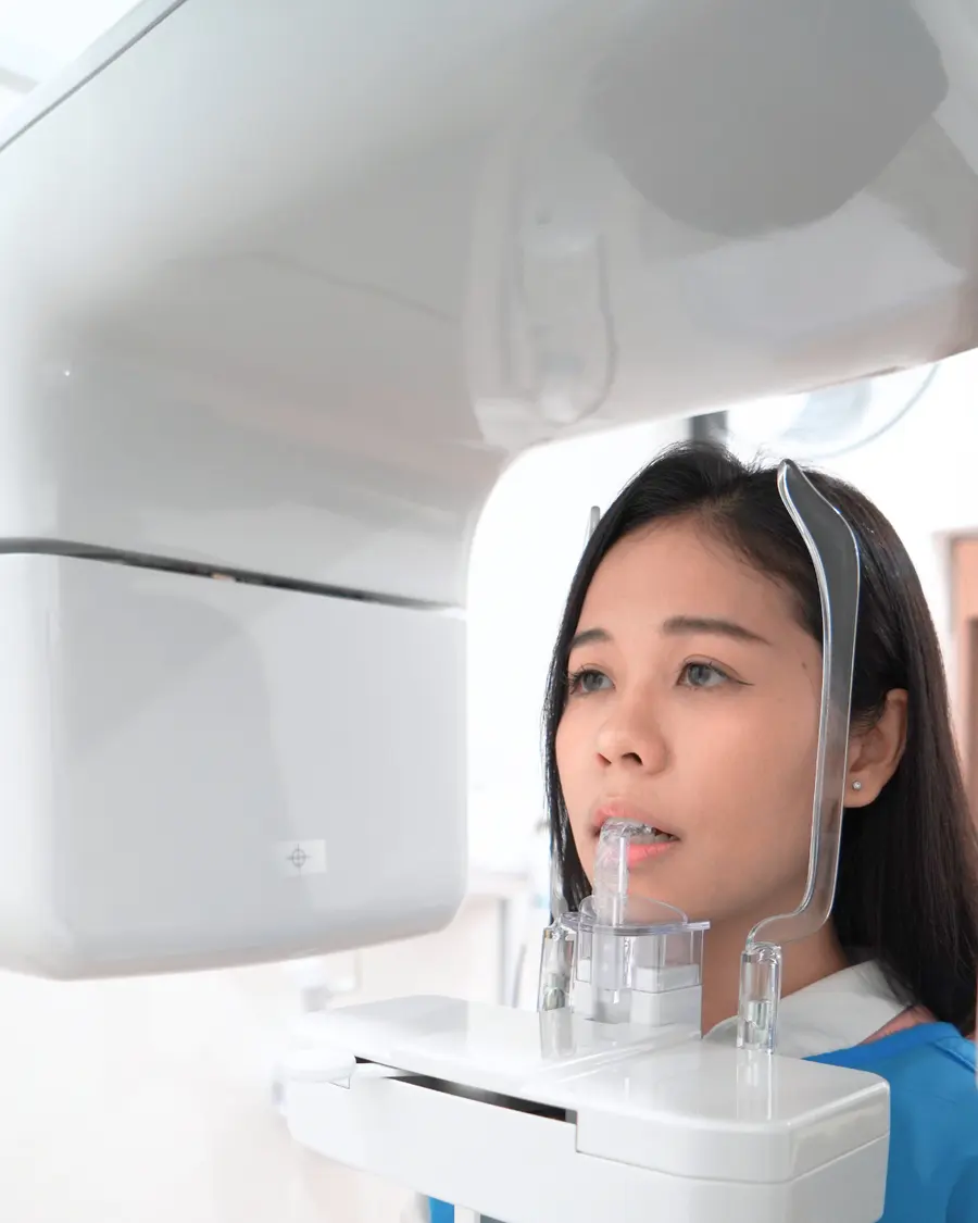
The dentist decides which image answers the question: a small periapical X-ray, bitewings, an OPG, a lateral cephalometric X-ray or a CBCT scan.
A small sensor is held in the mouth for intraoral X-rays. For an OPG, cephalometric or CBCT scan you stand or sit still at the machine for 10 to 40 seconds.
The exposure itself lasts a fraction of a second for an intraoral X-ray. An OPG or CBCT scan takes under a minute.
The digital image appears on screen immediately and is explained to you at the same visit.
Images are stored in your record and copies can be provided for another clinic or specialist.
The images belong to you. If you move clinics or are referred, ask us and we will send them across, which saves you both the dose and the fee of having them taken again.
The practical things patients ask about.
Tell us if you are or may be pregnant. A lead apron is used for intraoral images. Jewellery, glasses, hair clips and removable dentures come out for a panoramic or a CBCT, because metal shows on the image as a bright streak across whatever sits behind it.
A sensor is placed inside the mouth against the tooth and the exposure lasts a fraction of a second. The only uncomfortable part is holding the sensor still, and it is over before it becomes uncomfortable.
You stand or sit still with your chin on a rest while the arm rotates around your head, twenty seconds for an OPG and ten to forty for a CBCT. Nothing touches you and nothing is felt.
There is no afterwards. There is no residual radiation, nothing to avoid, no restriction on eating or driving, and no reason to keep away from children or anyone who is pregnant.
Tell us before rather than after if there is any chance you are pregnant. A dental X-ray directs a very small beam well away from the abdomen and is generally considered safe in pregnancy, and a non-urgent image can simply wait until afterwards, which is the easier decision for both of us.
Fees are quoted per case. These factors move the figure.
Periapical, bitewing, OPG, cephalometric or CBCT, and for CBCT the size of the area scanned.
Whether the image is part of a treatment plan such as implants or braces.
Copies of images on disc or by email for another provider.

Imaging fees are explained before the scan is taken. Images taken as part of a treatment plan are included in that plan's written quote.
Book a consultation or message us on WhatsApp. We explain your options and give a written plan before any treatment begins.
Each image answers a different question. The smallest one that answers yours is the one we take.
| Periapical | Panoramic OPG | 3D CBCT | |
|---|---|---|---|
| What it shows | One or two teeth in fine detail, from the crown to the root tip and the bone around it | Every tooth, both jaws, the sinuses and the jaw joints in a single image | Bone, nerves, sinuses and roots in three dimensions, viewable in any plane |
| Dimensions | Two dimensional | Two dimensional | Three dimensional |
| Usually taken for | Toothache, decay between teeth, checking a root canal, a suspected crack | A new patient overview, wisdom teeth, orthodontic planning, jaw pathology | Implant planning, surgical extractions near a nerve, complex root canal cases |
| Time it takes | A few seconds, with a small sensor inside the mouth | About twenty seconds standing still while the arm rotates | Ten to forty seconds, seated or standing |
| Dose in plain terms | Comparable to about a day of natural background radiation | Comparable to a few days | Days to a few weeks, depending on how large a field is scanned |
| What it cannot do | Shows only the area covered by the sensor | Structures overlap, so fine detail and thickness are lost | Not used routinely, because the dose is higher than a flat image |
A lateral cephalometric X-ray is the fourth type we take, used almost entirely for orthodontic planning, because it shows the relationship between the upper and lower jaws in profile. Recent images from another clinic can often be used instead of repeating one, so bring them or ask us to request them.
Open a topic for the full explanation.
X-rays are taken only when they change what we can diagnose or plan. Common reasons are a baseline at a new-patient examination, check-up images to look for decay between the teeth (usually every six months to two years depending on your risk), toothache or swelling, and planning before extractions, root canal treatment, braces or implants. They are also used to check wisdom teeth. Tell us if you are or may be pregnant, or if you have recent X-rays from another clinic that we can use instead.
Radiation dose is measured in microsieverts, which means nothing to most people, so the useful comparison is natural background radiation. Everyone receives roughly two to three millisieverts a year simply from the ground, the air and food, which works out at around six or seven microsieverts a day.
Against that, a digital intraoral X-ray is in the region of a day of background, a panoramic OPG a few days, and a cephalometric X-ray about a day. A dental CBCT covers a much wider range, from a few days for a small field looking at one implant site up to a few weeks for a large field of both jaws. For further context, a long haul flight delivers a few tens of microsieverts from cosmic radiation at altitude, and a medical CT scan of the head is roughly an order of magnitude above the largest dental CBCT.
None of this makes a dental X-ray free, and that is the point of taking one only when it answers a question that changes what we do. It does mean that the dose is not a reason to decline an image that would otherwise prevent a misdiagnosis.
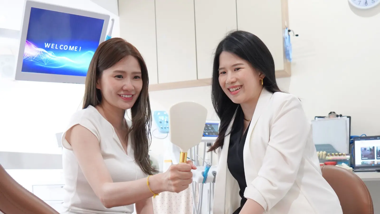
V Dental Clinic in SS2, Petaling Jaya provides dental x-rays with clear explanations, careful planning and follow-up at every step.
A detailed treatment plan and fee breakdown before treatment begins.
Strict sterilisation and hygiene protocols for every visit.
755+ Google reviews from our patients. Read them on Google.
Conveniently located in SS2, Petaling Jaya.
Before and after cases treated at our clinic, and what patients say on Google.
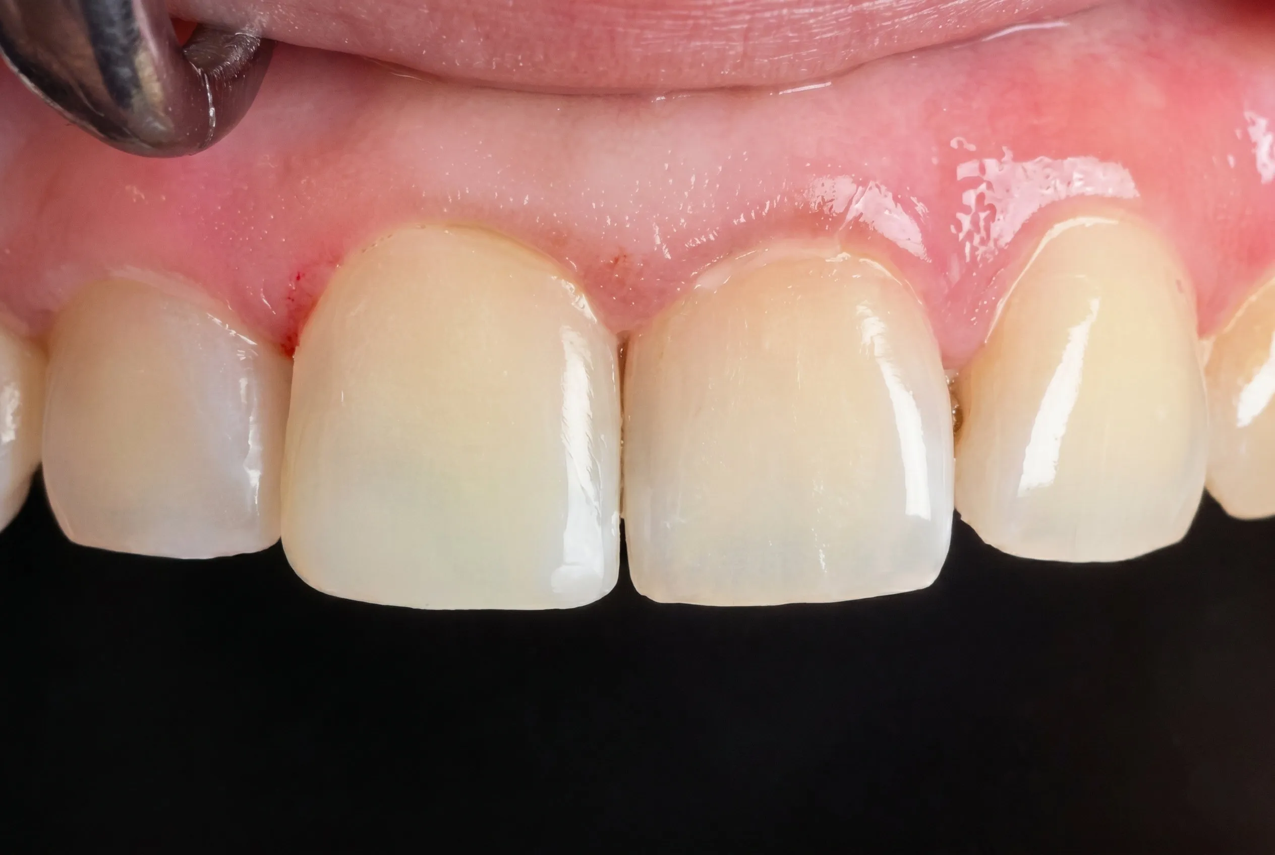
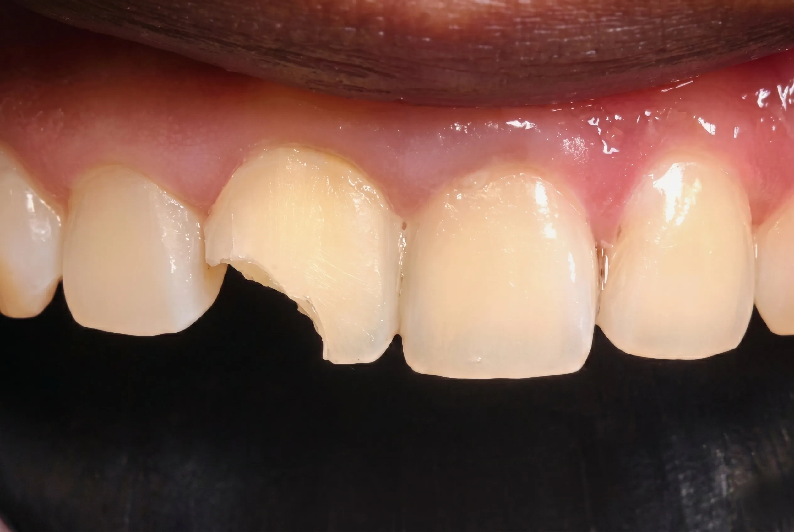
Restoring huge chipped tooth with aesthetic filling.
out of 5 on Google
Based on 755+ reviews
Adnan
July 2026
Thank you,Dr Koh, for the excellent dental implant treatment.The whole process was well explained,smooth and comfortable.Im very pleased with the results and would definitely recommend this clinic to others
View on GoogleSummer Lee
June 2026
The doctor was very good, patient, and professional. I went for braces treatment and had a great experience. The doctor explained everything clearly and made me feel comfortable throughout the consultation. Highly recommended!
View on GoogleNick Koay Han Yann
June 2026
Recommended by one of my relatives, Dr Koh is very friendly and informative. Did my scaling and filling. No issue, had a good experience.
View on GoogleEverything you need to know about this treatment.
Dental X-rays use very low doses, and digital sensors reduce them further. A single intraoral X-ray gives a dose comparable to a day or two of natural background radiation. A panoramic OPG is roughly equivalent to a few days, and a dental CBCT is higher but typically a fraction of a medical CT scan, depending on the size of the area scanned. We only take X-rays when they change the diagnosis or the plan.
An OPG is a single two-dimensional panoramic image of all the teeth and both jaws, used for general assessment, wisdom teeth and orthodontics. A CBCT is a three-dimensional scan that shows bone thickness, nerves and sinuses in any plane, used for implants, surgical extractions and complex root canal cases.
Before implant placement, when a wisdom tooth root lies close to the nerve, for root canal cases with unusual anatomy or persistent infection, for jaw joint problems, and when a cyst or lesion needs to be measured. Routine check-ups do not need a CBCT.
An intraoral X-ray takes a second, an OPG about 15 seconds and a CBCT under a minute. The images are digital and are on screen immediately, so the dentist can explain them at the same visit.
Yes, if they are recent, usually within six to twelve months, and of good quality. Bring the files or ask the clinic to email them to us. If the area of interest is not shown clearly, a new image may still be needed.
Dental X-rays give a very small dose directed away from the abdomen and are not ruled out in pregnancy, but we keep them to what is needed for diagnosis or to treat pain or infection, using the smallest possible field. Tell us if you are or may be pregnant before any X-ray.
Treatments that commonly come up alongside this one, either as an alternative or as the step before or after.
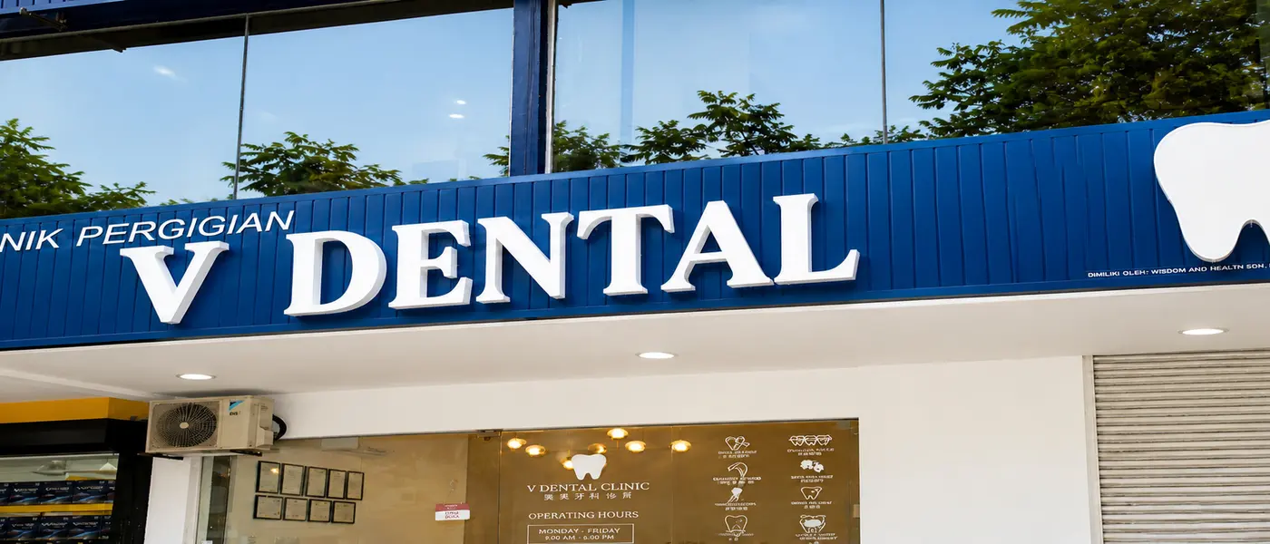
Walk-ins are welcome, but an appointment keeps your waiting time short.
Opening hours
Mon to Sat: 9:00 AM to 9:00 PM (MYT)
Sun: 9:00 AM to 6:00 PM (MYT)